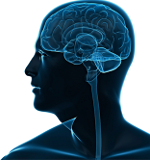
New Guidance on Assessing Neuroendocrine Dysfunction following TBI

In our May, 2014 post, we reported on research showing that traumatic brain injury, including mild traumatic brain injury (mTBI), can damage and cause dysfunction in the pituitary gland resulting in deficiencies in key hormones released by the pituitary gland, such as Growth Hormone (GH). As we explained in that post, the anatomy of the pituitary gland makes it particularly susceptible to the sheering injuries seen in TBI. These hormone deficiencies can produce many of the persistent symptoms seen following a TBI, such as fatigue, poor memory, depression, anxiety, emotional lability, exercise intolerance, lack of concentration and attention difficulties. (Although not always the case, these deficiencies can also produce physical symptoms, such as increased fat mass – especially in the abdominal area – and increased cholesterol.) We also noted findings showing that pituitary dysfunction can worsen over the five year period following an injury – in other words, that this is an issue that deserves to be monitored on an ongoing basis.
Subsequent studies have shown that the incidence of pituitary dysfunction in mTBI cases is higher in so-called “complicated mTBI” cases, with findings of skull fracture and/or intracranial abnormalities on imaging. In those cases, ongoing assessment is recommended, even where clinical manifestations are not clear, since those manifestations can be mild or subtle, leaving patients undiagnosed and untreated without neuroendocrine screening.
Standard practice may not be accurate in diagnosing Growth Hormone deficiency
The standard practice for assessing Growth Hormone (GH) deficiencies is the “ICF-1” test, which measures “insulin-like growth factor-1” in the blood as a proxy for GH. A recent study in the Journal of Neurotrauma notes that 50% of adults with GH deficiency have IGF-1 levels within the normal reference range. The authors also note that “direct serum assessment is unreliable because of the pulsatile release of GH and results in serum fluxuations within a 24-h period.”
“To definitively diagnose” GH-deficiency, the authors conclude, “provocative testing is essential.” (Provocative testing involves measuring the body’s response to some substance.) The two effective provocative tests recommended are the “insulin tolerance test” (ITT) and the “glucagon stimulation test” (GST). The insulin test is considered the “gold standard” but cannot safely be performed on patients with seizures, so may not be appropriate for many patients with TBI. In those cases, the GST is preferred. The researchers found, as expected, that TBI patients with GH deficiency had more disability than patients without this condition. They also found that patients with GH deficiency had higher levels of depressive symptoms and lower levels of testosterone.
As the authors note, other studies have found that GH replacement in deficient patients often leads to improvement in symptoms. Undiagnosed pituitary deficiency therefore has the potential to impede rehabilitation and recovery following a TBI, especially in the chronic phase of recovery. Patients who receive testing should inquire about the testing methodology to insure that accurate provocative testing is used.